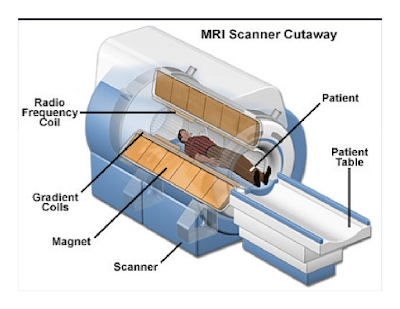
MRl is a diagnostic technique that generates images of the internal body. It specilizes in creating a thin-section image of any part of the body-especially the heart, veins, arteries, brain 🧠, and central nervous system-from any angle or direction.
This MRl scan of a normal head shows the brain, airways, any soft tissues of the face. The large cerebral cortex, appearing in white and black, from the bulk of the brain 🧠 tissue; the circular cerebellum, center, in light black, are also prominently shown.
Magnetic Resonance Imaging (MRI), medical, diagnostic technique that combines strong magnetic field, radio waves, and computer technology to create images of the body using the principles of nuclear magnetic resonance.
A versatile, powerful, and sensitive tool, MRI can generate thin-section computerized images of any part of the body - including the heart, arteries and veins-from any angle direction, without surgical invasion and in a relatively short period of time.
MRI also create "maps" of bio-chemical and anotomical information that provides new knowledge and may allow early diagnosis of many diseases. In 2003 Paul Lauterbur of the United States and Sir Perter Mansfield and United Kingdom shared the Nobel Prize in physiology medicine for their contribution of MRI technology.
MRI is possible in human body because the body is filled with small biological "magnets," the most abundant and responsive of which is the proton, the nucleus of the hydrogen atom. The principles of MRI take advantage of the random distribution of protons, which possess fundamental magnetic properties. One the patient is placed in the cylindrical magnet, the diagnostic process follows three bases steps.
First, MRI creates a study state within the body by placing the body in a steady magnetic field that is 30,000 times stronger than Earth's magnetic field. Then MRI stimulates the body with radio waves to change the steady -state orientation of protons. It then stops the radio waves and "listens" to the body's electromagnetic transformations at a selected frequency. The transmitted signal is used to construct internal images of the body using principles similar to those developed for computerized axial tomography, or CAT scanners.
In current medical practice, MRI is preferred for diagnosing most diseases of the brain 🧠 and central nervous system. MRI scanners provide equivalent resolution and superior contrast resolution to that of X-Ray CAT scanners MRI scanners also provide imaging complementary to X-Ray images because MRI can distinguish soft tissues in the both normal diseased states.
Base Peak. The set of ions produced from a molecule can be analyzed since each has its own m/e ratio, and produces a signal whose intensity is due to the relative abundance of that ion. The largest peak found in a mass spectrum (that of highest intersity) is called the base peak, and is given the numerical value of 100. The spectrum is highly characteristics of a particular compound.
Molecular Ion: The ion formed by removing one electron from the patent molecule is called molecular ions or parent ion. The molecular Ion peak is usually represented as M+. It may or may not be the peak of highest intersity. The molecular ions is the most important ion, since it's mass is the Molecular Weigh of the parent molecule.
The use of mass spectroscopy is not limited to the determination of the molecular weight of compounds. It also has great untlity in the elucidation of the structure of a molecule. The fragmentation process of organic molecules follow certain patterns in that the fragments broken off are related to bond strengths and the stability of the species formed. Since certain structural features in molecule produce definite characteristics fragmentation patterns, the identification of these fragments amd their relatives intensities in a mass spectrum can be used to determine the structure of organic compound.


